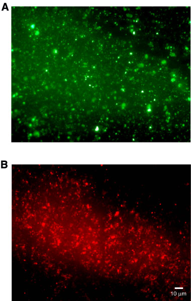
Figure F18. Photomicrograph showing bacterial cells from an upper sediment layer from a depth of 1.45 mbsf (Sample
200-1224C-1H-4, 145-150 cm). A. After staining with the DNA-binding fluorochrome SYBR Green I. B. Hybridization with the Bacteria-specific, CY3-labeled probe EUB338. Note the numerous bacteria responding to the specific hybridization, indicating the high amount of metabolically active bacteria within the sediment.